What Part of Synovial Joints is Continuous With the Periosteum
Introduction [edit | edit source]
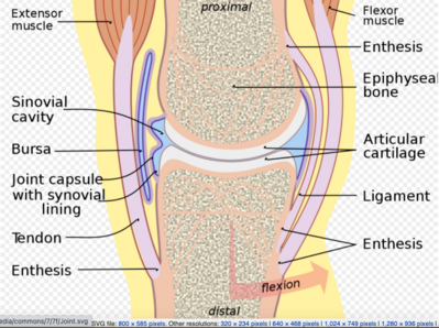
The three joints in the body (Histologically) are fibrous, cartilaginous, and synovial.
Synovial joints are the most common type of joint in the body (see image 1). These joints are termed diarthroses, meaning they are freely mobile.[1]
A key structural characteristic for a synovial joint that is not seen at fibrous or cartilaginous joints is the presence of a joint cavity. The joint cavity contains synovial fluid, secreted by the synovial membrane (synovium), which lines the articular capsule. This fluid-filled space is the site at which the articulating surfaces of the bones contact each other. Hyaline cartilage forms the articular cartilage, covering the entire articulating surface of each bone. The articular cartilage and the synovial membrane are continuous. A few synovial joints of the body have a fibrocartilage structure located between the articulating bones. This is called an articular disc, which is generally small and oval-shaped, or a meniscus, which is larger and C-shaped.[2] [3].
Synovial joints are often further classified by the type of movements they permit. There are six such classifications: hinge (elbow), saddle (carpometacarpal joint), planar (acromioclavicular joint), pivot (atlantoaxial joint), condyloid (metacarpophalangeal joint), and ball and socket (hip joint).[2]
Features of all Synovial Joints [edit | edit source]
- Articular capsule with synovial membrane
- Synovial cavity containing synovial fluid
- Hyaline articular cartilage: acts like a Teflon® coating over the bone surface, allowing the articulating bones to move smoothly against each other without damaging the underlying bone tissue.[3] [1]
Additional features within some Synovial Joints [edit | edit source]
- Fibrocartilage structure located between the articulating bones: Articular disc, which is generally small and oval-shaped, or a Meniscus, which is larger and C-shaped. These structures can serve several functions, depending on the specific joint. Can serve to smooth the movements between the articulating bones eg at the temporomandibular joint.
- Intrinsic ligament: fused to or incorporated into the wall of the articular capsule
- Intracapsular ligament: located inside of the articular capsule.
- Intra-capsular tendons eg. popliteus tendon within the knee joint
- Intra-articular tendons eg. long head of biceps tendon within the shoulder joint[1]
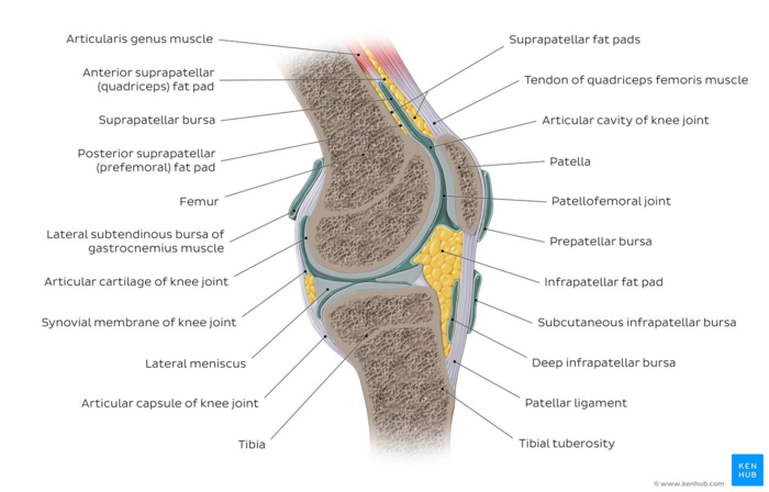
Image 2: Knee joint. In a Synovial joint, the ends of bones are encased in smooth cartilage. Together, they are protected by a joint capsule lined with a synovial membrane that produces synovial fluid. The capsule and fluid protect the cartilage, muscles, and connective tissues. [4]
Additional features surrounding some Synovial Joints [edit | edit source]
-
Fat pads: serve as a cushion between the bones.[3]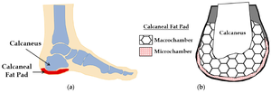
- Bursae: small fluid-filled sacs lined by a synovial membrane with an inner capillary layer of synovial fluid. It provides a cushion between bones and tendons and/or muscles around a joint. This helps to reduce friction between the bones and allows free movement. They may or may not communicate with the joint space.
- Extra-capsular ligaments: strong bands of fibrous connective tissue. These strengthen and support the joint by anchoring the bones together and preventing their separation. Ligaments allow for normal movements at a joint, but limit the range of these motions, thus preventing excessive or abnormal joint movements. [3]
- Tendons
Sesamoid bones: focal areas of ossification within tendons as they pass over joints. They can also occur in ligaments and usually measure a few millimetres in diameter. Their function is purported to be to alter the direction of the tendon and modify pressure, thereby reducing friction[1].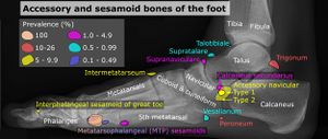
Additional Support [edit | edit source]
At many synovial joints, additional support is provided by the muscles and their tendons that act across the joint.
- Tendons are dense connective tissue structure attaching a muscle to bone. As forces acting on a joint increase, the body will automatically increase the overall strength of contraction of the muscles crossing that joint, thus allowing the muscle and its tendon to serve as a "dynamic ligament" to resist forces and support the joint. This type of indirect support by muscles is very important at the shoulder joint, for example, where the ligaments are relatively weak.
Types of Synovial Joints [edit | edit source]
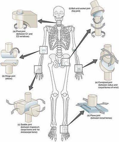
Image 4: Types of Synovial Joints: The six types of synovial joints allow the body to move in a variety of ways. (a) Pivot joints allow for rotation around an axis, such as between the first and second cervical vertebrae, which allows for side-to-side rotation of the head. (b) The hinge joint of the elbow works like a door hinge. (c) The articulation between the trapezium carpal bone and the first metacarpal bone at the base of the thumb is a saddle joint. (d) Plane joints, such as those between the tarsal bones of the foot, allow for limited gliding movements between bones. (e) The radiocarpal joint of the wrist is a condyloid joint. (f) The hip and shoulder joints are the only ball-and-socket joints of the body.
The six types of synovial joints are:
- Plane Joints: Multiaxial joint , the articular surfaces are essentially flat, and they allow only short nonaxial gliding movements. Examples are the gliding joints introduced earlier—the intercarpal and intertarsal joints, and the joints between vertebral articular processes. Gliding does not involve rotation around any axis, and gliding joints are the only examples of nonaxial plane joints
- Hinge Joints: Uniaxial Joint, the cylindrical end of one bone conforms to a trough-shaped surface on another. Motion is along a single plane and resembles that of a mechanical hinge. Uniaxial hinge joints permit flexion and extension only, typified by bending and straightening the elbow and interphalangeal joints.
- Pivot Joints: Uniaxial Joint , the rounded end of one bone conforms to a "sleeve" r ring composed of bone (and possibly ligaments) of another. The only movement allowed is uniaxial rotation of one bone around its own long axis. An example is the joint between the atlas and dens of the axis, which allows you to move your head from side to side to indicate "no." Another is the proximal radioulnar joint, where the head of the radius rotates within a ringlike ligament secured to the ulna.
- Condyloid ( ellipsoidal ) Joints: Biaxial joints , The oval articular surface of one bone fits into a complementary depression in another . The important characteristic is that both articulating surfaces are oval. The biaxial condyloid joints permit all angular motions, that is, flexion and extension, abduction and adduction, and circumduction. The radiocarpal (wrist) joints and the metacarpophalangeal (knuckle) joints are typical condyloid joints.
- Saddle Joints: Biaxial Joints , resemble condyloid joints, but they allow greater freedom of movement. Each articular surface has both concave and convex areas; that is, it is shaped like a saddle. The articular surfaces then fit together, concave to convex surfaces. The most clear-cut examples of saddle joints in the body are the carpometacarpal joints of the thumbs, and the movements allowed by these joints are clearly demonstrated by twiddling your thumbs.
- Ball-and-Socket Joints: Multiaxial joint , In ball-and-socket joints , the sherical or hemispherical head of one bone articulates with the cuplike socket of another. These joints are multiaxial and the most freely moving synovial joints. Universal movement is allowed (that is, in all axes and planes, including rotation). The shoulder and hip joints are the only examples. [2]
Nerve Supply [edit | edit source]
Sensory and autonomic fibers innervate synovial joints:
- The autonomic nerves are vasomotor in function, controlling the dilation or constriction of blood vessels.
- The sensory nerves of the articular capsule and ligaments (articular nerves) provide proprioceptive feedback from Ruffini endings and Pacinian corpuscles. Proprioception of the joint permits reflex control of posture, locomotion, and movement. Free nerve endings convey pain sensation that is diffuse and poorly localized. The articular cartilage has no nerve supply.
Two general principles apply to synovial joint innervation:
- Hilton's law states: Articular nerves supplying a joint are branches of the nerves that supply the muscles responsible for moving that joint. Hence irritation of articular nerves causes a reflex spasm of the muscles which position the joint for the greatest comfort. These nerves also supply the overlying skin, providing a mechanism for referred pain from joint to skin.
- Gardner's observation: When part of the articular capsule that is tightened, by contraction of a group of muscles, they receives nerve supply from the same nerves that innervate the antagonist muscles. This relationship provides local reflex arcs that stabilize the joint[2].
Blood Supply [edit | edit source]
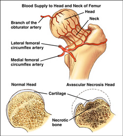
Synovial joints receive vascular supply through a rich anastomosis of arteries extending from either side of the joint ie the periarticular plexus. Some vessels penetrate the fibrous capsule to form a rich plexus deeper in the synovial membrane.
The articular cartilage, which is avascular hyaline cartilage, is nourished by the synovial fluid.
Lymphatic vessels for every joint follow the lymph drainage of the surrounding tissue—some joints house lymph nodes, like the popliteal lymph nodes in the popliteal fossa of the knee[1].
[edit | edit source]

Typically seen in patients with osteoarthritis, rheumatoid arthritis, and other arthritic conditions. Joints contain sensory nerves called baro-receptors which respond to changes in atmospheric pressure. These receptors especially react when there is low barometric pressure, meaning the atmosphere has gone from dry to moist, like when it is going to rain.
When pressure in the environment changes, we know that the amount of fluid in the joint or the pressure inside the joint fluctuates with it. Individuals with arthritic joints feel these changes much more because they have less cartilage to provide cushioning.[5]
References [edit | edit source]
- ↑ 1.0 1.1 1.2 1.3 1.4 Radiopedia Synovial Joints Available: https://radiopaedia.org/articles/synovial-joints(accessed 20.6.2021)
- ↑ 2.0 2.1 2.2 2.3 Juneja P, Hubbard JB. Anatomy, Joints. 2018 Available: https://www.ncbi.nlm.nih.gov/books/NBK507893/ (accessed 20.6.2021)
- ↑ 3.0 3.1 3.2 3.3 OSE Synovial joints Available:https://open.oregonstate.education/aandp/chapter/9-4-synovial-joints/ (accessed 20.6.2021)
- ↑ Knee joint image - © Kenhub https://www.kenhub.com/en/library/anatomy/the-knee-joint
- ↑ Thomas Jefferson University Hospital. "People With Joint Pain Can Really Forecast Thunderstorms." ScienceDaily. ScienceDaily, 3 June 2008. Available:https://www.sciencedaily.com/releases/2008/05/080530174619.htm (accessed 20.6.2021)
Source: https://www.physio-pedia.com/Synovial_Joints
Posting Komentar untuk "What Part of Synovial Joints is Continuous With the Periosteum"